
If you’re new to brain CTs, here are some quick points to keep in mind about normal scans and what they show.
Key Takeaways
- A normal brain CT shows no signs of bleeding, swelling, or obvious injury.
- The ventricles and brain structures should be even and in the right place.
- Bone around the brain looks solid, with no cracks or breaks.
- Doctors look for symmetry and clear borders between gray and white matter.
- A normal scan rules out things like large tumors, big bleeds, or major fractures.
Understanding A Normal Brain CT
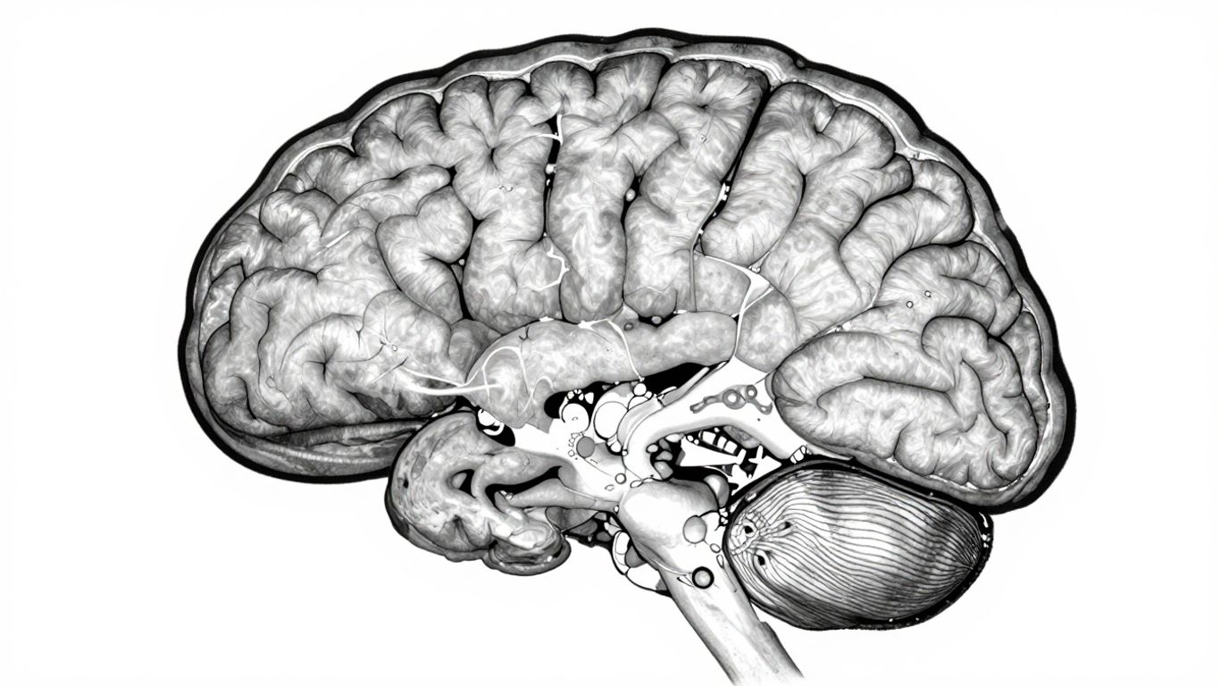
So, you're getting a brain CT scan, or maybe you're just curious about what doctors look for. It's a pretty common imaging test, and understanding what a normal scan looks like can be really helpful. Think of it like looking at a map – you need to know what the usual landmarks are before you can spot anything out of the ordinary.
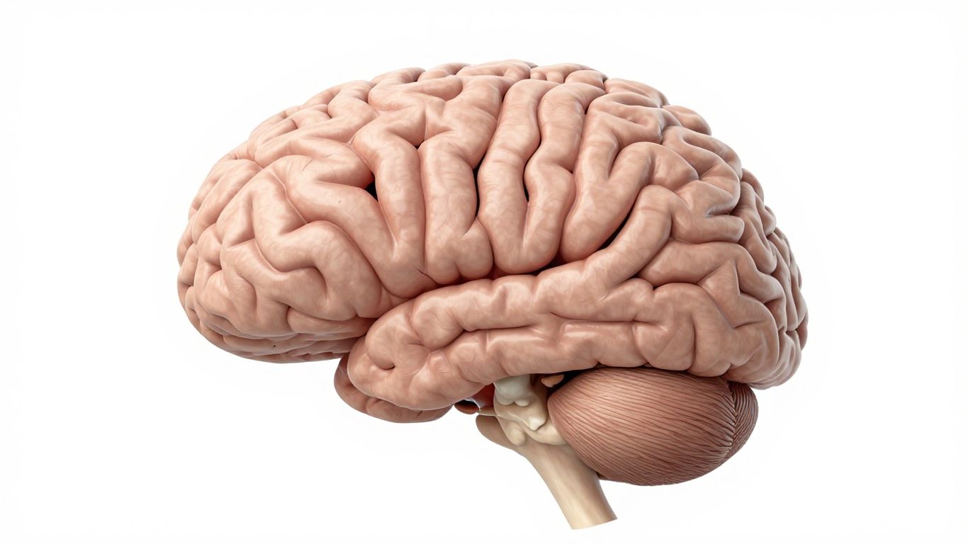
What Is A Brain CT Scan?
A CT scan, which stands for computed tomography, uses X-rays to create detailed cross-sectional images, or 'slices,' of your brain. It's a quick and painless way to get a look inside your head. The machine takes multiple X-ray images from different angles, and a computer puts them together to form these detailed pictures (per the NIH). Sometimes, a special dye called contrast is used to make certain parts, like blood vessels, stand out more clearly. This is usually given through an IV (per MedlinePlus).
How A Normal Brain CT Appears
On a normal CT scan, the brain tissue itself will have a relatively uniform grey appearance. Different parts of the brain have slightly different shades, and that's normal. The key is that there aren't any sudden, bright white spots (which can indicate fresh blood or calcification) or dark black areas (which might suggest swelling or a stroke) where they shouldn't be (per MedlinePlus). The fluid-filled spaces, called ventricles, should look dark and be a normal size. Bone, like your skull, will appear bright white because it's dense. The goal is to see clear boundaries between these different tissues.
Key Structures To Examine On A Normal Brain CT
When a radiologist looks at a brain CT, they're checking several things. They want to make sure:
- The brain is symmetrical: Both sides of the brain should look pretty much the same. This includes the grooves (sulci) and ridges (gyri) on the surface.
- Grey-white differentiation is clear: Normally, you can easily tell the difference between the grey matter (the outer part of the brain) and the white matter (the inner part). This distinction is important.
- The ventricles are normal: These are the fluid-filled cavities within the brain. They shouldn't be too large (dilated) or squashed (compressed). A normal size and shape are what we expect.
- The cisterns are open: These are spaces around the brain that contain cerebrospinal fluid. They should be clear and not filled with anything abnormal, like blood.
When you look at a CT scan, remember that the right side of the image usually shows the left side of the patient's brain, and vice versa. It's like looking at someone in a mirror. Also, different settings, called 'windows,' are used to best see different tissues – one for the brain itself and another for the bone.
It's important to remember that a CT scan is just one piece of the puzzle when it comes to understanding brain health. For instance, tests like nerve conduction studies can give different kinds of information about nerve function. But for a quick look at potential bleeding or major structural issues, a CT is often the first step.
Systematic Approach To Reading A Brain CT
So, you've got a brain CT scan, and now what? It's not just about looking at a bunch of black and white slices. To really get a handle on what's going on, you need a method. Think of it like following a recipe – skip a step, and things might not turn out right. A systematic approach helps make sure you don't miss anything important, especially when time is of the essence. It's about being thorough and organized, so you can spot potential issues quickly and accurately. This methodical way of looking at the scans is key to interpreting them correctly.
Evaluating For Bleeding
This is usually the first thing on anyone's mind when looking at a brain CT, and for good reason. Bleeding inside the skull can be a serious emergency. You're looking for areas that appear brighter than the surrounding brain tissue. These bright spots, or hyperdensities, can indicate fresh blood. It's important to know where to look for different types of bleeds, which have characteristic appearances on CT (per NIH):
- Epidural hematoma: Often lens-shaped, doesn't cross suture lines.
- Subdural hematoma: Crescent-shaped, can cross suture lines.
- Intraparenchymal hemorrhage: Bleeding within the brain tissue itself, often in specific areas like the basal ganglia if related to high blood pressure.
- Subarachnoid hemorrhage: Blood in the space around the brain, frequently from a ruptured aneurysm.
- Intraventricular hemorrhage: Blood within the brain's fluid-filled ventricles.
The mnemonic "Blood Can Be Very Bad" is a helpful reminder to always check for bleeding first.
Assessing The Cisterns
Next up, we check the cisterns. These are fluid-filled spaces in the brain's subarachnoid space. They're like little reservoirs of cerebrospinal fluid. When you look at a normal CT, these spaces should be open and clear. You're essentially asking two main questions about the key cisterns (like the circummesencephalic, suprasellar, quadrigeminal, and Sylvian cisterns):
- Is there any blood present in these spaces?
- Are the cisterns open and not compressed?
If these spaces are squeezed or filled with blood, it can signal increased pressure inside the skull or bleeding, respectively. Keeping these spaces clear is vital for proper brain function.
Examining Brain Symmetry And Differentiation
Finally, you'll want to look closely at the brain tissue itself. Pay attention to symmetry – does one side look like the other? The grooves (sulci) and ridges (gyri) on the surface should appear similar on both the left and right sides. Also, check for grey-white differentiation. Normally, you can clearly see the boundary between the grey matter (outer layer) and the white matter (inner part). If this boundary starts to blur or disappear, it can be an early sign of a problem, like a stroke (per NIH). You'll also want to see if the brain structures are midline and if the ventricles (the fluid-filled cavities) look normal in size and shape. Any shift or compression of these structures can indicate a significant issue. This systematic review helps ensure that you're not overlooking subtle but important changes on the scan, similar to how a systematic approach to interpreting neck CT findings is vital for accurate diagnosis.
Interpreting Brain CT Findings
So, you've got a brain CT scan report, and now you're trying to make sense of it all. It can feel like deciphering a secret code sometimes, but understanding a few key things can make a big difference. We're going to break down how to look at those slices and what the different shades of gray actually mean.
Understanding Hyperdense and Hypodense Areas
When you look at a CT scan, you'll notice different shades of gray. These shades tell us about the density of the tissue. Think of it like this: denser things show up brighter, and less dense things show up darker.
- Hyperdense areas appear bright white. This usually means something is more dense than the surrounding brain tissue. Common causes include fresh blood (hemorrhage), calcium deposits, or contrast dye used during the scan. Spotting a hyperdense area is often the first clue that something acute might be happening.
- Hypodense areas appear darker. This suggests the tissue is less dense. This can be seen in areas of swelling (edema), old damage, or sometimes in tumors or areas affected by a stroke where tissue has died.
It's important to remember that these terms just describe what the scan shows, not necessarily the cause. A radiologist puts these findings together with your symptoms to figure out what's going on.
Assessing Ventricles for Dilation or Compression
Inside your brain, there are fluid-filled spaces called ventricles. These spaces contain cerebrospinal fluid (CSF), which acts as a cushion and helps remove waste. On a CT scan, these ventricles normally look dark because the fluid is less dense than brain tissue.
- Dilation: If the ventricles look larger than they should, it's called dilation. This can happen if there's a blockage somewhere in the CSF pathway, causing the fluid to build up and push outwards. This buildup increases pressure inside the skull, a condition known as hydrocephalus. Sometimes, even without a blockage, the brain tissue itself might shrink, making the ventricles appear larger by comparison.
- Compression: The opposite can also happen. If there's something pushing on the ventricles, like a tumor or significant swelling from an injury, they can appear squashed or compressed. This compression can also cause a shift in the brain's midline, which is a serious sign.
Identifying Bone and Skull Integrity
While the CT scan's main focus is often the brain tissue, it also gives a really good look at the bones of the skull. This is especially important if there's been any kind of trauma.
- Fractures: The scan can clearly show if there are any breaks or cracks in the skull bones. Even small fractures can sometimes be missed on regular X-rays, but CT is much better at picking them up. You'll see these as distinct breaks in the white bone lines.
- Other Bony Abnormalities: Sometimes, CT can also show other issues with the bone, like thickening, thinning, or signs of old injuries.
When looking at these findings, it's always about putting the pieces together. A dark spot might be a stroke, or it might just be a normal part of aging. That's why a trained professional is key to interpreting the full picture.
It's easy to get lost in the details of a CT scan, but remember, the goal is to see if there are any immediate dangers like bleeding or significant swelling. The scan is a snapshot, and it's the radiologist's job to interpret that snapshot in the context of why the scan was ordered in the first place. They're looking for things that are out of the ordinary and could be causing problems right now.
When A Brain CT Is Indicated
Knowing when to get a brain CT scan can definitely feel overwhelming, especially if you're worried about new symptoms or a recent accident. Doctors don’t usually order these scans unless there’s a real reason to check for problems with the brain or the surrounding tissue. Here’s a breakdown of some of the most common reasons:
Traumatic Brain Injuries And Head Trauma
- Any blow to the head—from car crashes, sports, or a simple fall—could cause problems you can’t see right away.
- A CT scan is often the first tool used to look for things like skull fractures, brain bleeding, or swelling (per the NIH). Fast answers can be critical here.
- It also helps doctors decide if surgery or close monitoring is needed.
| Injury Type | Why CT May Be Needed |
|---|---|
| Falls/accidents | Check for fractures, bleeding, swelling |
| Sports injuries | Rule out concussion complications |
| Assault or trauma | Detect hidden brain injury |
If you hit your head and notice things like confusion, vomiting, or passing out—even if you seem fine at first—a scan might be recommended to cover all the bases.
Symptoms Suggesting Stroke Or Bleeding
- Sudden numbness, weakness (especially on one side), confusion, or trouble speaking/walking are all red flags.
- Brain CTs can show if a stroke is caused by a clot or a bleed, and help doctors act quickly.
- Around suspected stroke, CT is used right away to rule out other causes and spot bleeds that need urgent attention (per the Mayo Clinic).
Odd symptoms after a head injury or any sudden change—like confusion, memory loss, or weakness—shouldn’t be ignored, because a CT could spot a treatable cause before it gets worse.
Evaluating For Tumors Or Lesions
- Persistent headaches, vision issues, or unexplained seizures sometimes trigger the need for a brain scan.
- A CT can find masses, growths, or swelling that might not show up with just exams and blood work.
- It is also useful if you have a known cancer that could spread to the brain, or if a new neurological symptom starts and the cause is unclear.
Sometimes, your doctor will also use a CT scan if other tests or exams point to an unexplained problem—and in those cases, it can be helpful for guiding next steps. For more details on how brain scans fit into a complete workup, check out expert care for brain and nerve health.
In short, a brain CT scan is not an everyday test—there’s always a specific reason for ordering it, usually with the goal of finding fast answers to urgent or puzzling issues.
Comparing CT To Other Imaging Modalities
When we talk about looking inside the brain, CT scans are a common tool, but they're not the only game in town. It's helpful to know how they stack up against other methods, like MRI, to understand what each is best for.
CT Versus MRI For Brain Conditions
Think of a CT scan as a really fast, detailed X-ray. It's great for seeing bone and spotting quick issues like bleeding or major trauma. CT scans are often the first choice in emergency situations because they are quick and widely available. They can show us things like skull fractures or large hemorrhages pretty clearly. However, when it comes to the nitty-gritty details of soft tissues, like the brain itself, CT isn't as sensitive as MRI. It can miss subtle changes or inflammation.
MRI, on the other hand, uses magnetic fields and radio waves to create incredibly detailed images of soft tissues. It's like going from a black-and-white photo to a high-definition color movie for the brain. MRI is much better at showing things like tumors, inflammation, multiple sclerosis (MS) plaques, and smaller strokes. The trade-off? MRI scans take longer, can be noisy, and aren't suitable for everyone (like those with certain metal implants) (per the NIH). If you're trying to figure out the exact cause of subtle cognitive changes, an MRI might give you more information than a CT.
Limitations Of CT In Detecting Certain Pathologies
While CT is a workhorse, it has its blind spots. It's not the best tool for seeing inflammation in the membranes covering the brain, for instance. Subtle changes in brain tissue, like early signs of certain degenerative diseases or small areas of inflammation, can easily be missed on a CT. If a doctor suspects something like meningitis or very early-stage tumors, they'll likely lean towards an MRI. CT also has a harder time differentiating between different types of soft tissue compared to MRI.
Role Of CT In Guiding Procedures
Despite its limitations for certain diagnoses, CT plays a vital role in guiding medical procedures. Because it's fast and good at showing spatial relationships, it's often used in real-time during surgeries or interventions. For example, a neurosurgeon might use CT guidance to precisely place a needle or instrument during a biopsy or to drain fluid. It's also invaluable in the emergency room for quickly assessing the extent of injuries after an accident, helping doctors decide on the immediate course of action. For conditions like Transient Global Amnesia (TGA), a CT scan is often one of the initial tests used to rule out more serious causes like stroke or bleeding, helping to confirm the diagnosis.
What A Normal Brain CT Rules Out
So, you've had a brain CT, and the radiologist says it looks normal. That's usually great news! But what does 'normal' actually mean in this context? A normal CT scan is a powerful tool because it helps rule out a lot of serious, immediate problems that could be happening in your head. It's like a quick check to make sure the big, scary stuff isn't there.
Absence of Acute Bleeding
One of the most critical things a CT scan can quickly identify is bleeding within the brain. This includes conditions like a hemorrhagic stroke, where a blood vessel bursts, or bleeding from a head injury. Blood shows up as a bright white area, called hyperdense, on a CT scan. If your scan is clear of these bright spots in the brain tissue or the spaces around it, it means there's no evidence of acute bleeding. This is a huge relief, especially if you came in with symptoms like a sudden, severe headache or after a fall. It doesn't necessarily mean there are no issues, but it rules out the most life-threatening bleeding events that need immediate intervention. For example, a CT scan is often the first imaging procedure done after symptoms of a stroke appear, helping to confirm the diagnosis and rule out other conditions.
Normal Ventricular Size and Position
The ventricles are fluid-filled spaces within the brain. In a normal CT, these ventricles should be a standard size and located centrally. If there's swelling, a blockage, or a mass, these ventricles can become enlarged (dilated) or pushed out of place (shifted). A normal scan shows these structures looking just as they should – not too big, not too small, and right where they belong. This absence of ventricular dilation or compression suggests there isn't significant pressure building up inside the skull from things like hydrocephalus or a large tumor.
No Obvious Fractures or Mass Lesions
When looking at the skull itself, a normal CT scan will show intact bone without any breaks or fractures. This is especially important if you've experienced head trauma. The scan also checks for obvious mass lesions, which could be tumors or other abnormal growths. While CT isn't as detailed as MRI for soft tissues, it's excellent at spotting larger masses or significant structural abnormalities. So, a normal result means the bones of your skull look solid, and there aren't any large, suspicious masses readily visible within the brain tissue on this particular scan.
It's important to remember that a 'normal' CT scan is a snapshot in time. It effectively rules out many urgent conditions, but it doesn't necessarily explain every single symptom you might be experiencing. Sometimes, subtle changes or conditions that don't show up well on CT might still be present. Always discuss the full results and any lingering concerns with your doctor.
While a CT scan is great for spotting acute issues like bleeding or fractures, it has limitations. For instance, it's not as sensitive as MRI for detecting inflammation or subtle soft tissue changes. This is why, depending on your symptoms and medical history, your doctor might recommend other imaging tests, like an MRI, for a more detailed look.
Conclusion
Understanding a normal brain CT doesn’t have to be scary or confusing. With a little practice, you can get familiar with what a healthy scan looks like and what doctors are checking for. If you ever have questions about your own scan, always ask your healthcare provider—they’re there to help you make sense of the results. Remember, a normal brain CT is just one piece of the puzzle when it comes to your brain health.
Frequently Asked Questions
What is a brain CT scan?
A brain CT scan is a special X-ray that takes pictures of your brain from different angles. It helps doctors see inside your head without surgery.
What does a normal brain CT look like?
On a normal brain CT, the brain looks even on both sides, with no bright or dark spots that shouldn't be there. The spaces in the brain (ventricles) are the right size, and the skull is not broken.
Why would someone need a brain CT?
Doctors order a brain CT if you’ve had a head injury, bad headache, stroke symptoms, or if they think you might have a tumor or infection.
Can a normal brain CT rule out all problems?
No, a normal brain CT can’t catch every problem. Some small strokes, infections, or early disease changes might not show up. That’s why doctors use other tests if they need more answers.
How is a CT scan different from an MRI?
CT scans are faster and better for finding bleeding or bone breaks. MRIs take longer but can show more detail, especially for soft tissue and some diseases.
Is a brain CT safe?
Brain CTs use a small amount of radiation, but for most people, the health benefits outweigh the small potential risk (per the FDA). If you’re pregnant or have kidney problems, let your doctor know before the scan.
Sources
- National Institute of Biomedical Imaging and Bioengineering – Computed Tomography (CT)
- MedlinePlus – Head CT scan
- National Institute of Neurological Disorders and Stroke – Traumatic Brain Injury
- Mayo Clinic – Stroke: Diagnosis & treatment
- National Institute of Biomedical Imaging and Bioengineering – Magnetic Resonance Imaging (MRI)
- U.S. Food & Drug Administration – Computed Tomography (CT) Scans and Cancer